SRH Workflow
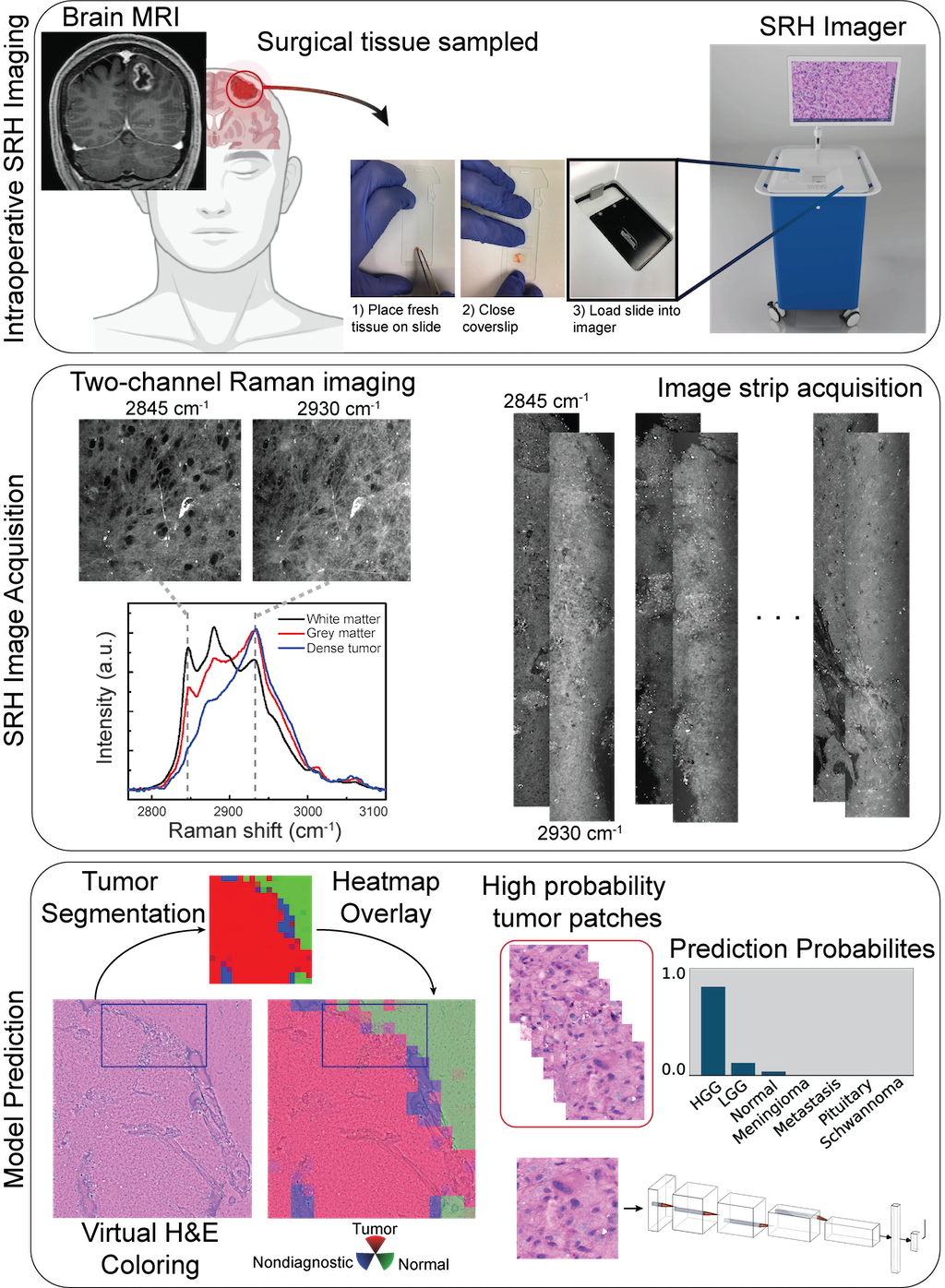
A patient with a newly diagnosed brain lesion undergoes a surgery for tissue diagnosis and/or tumor resection. The tumor specimen is sampled from the patient's tumor and directly loaded into a premade, disposable microscope slide. The specimen is placed into the SRH imager for rapid optical imaging. SRH images are acquired sequentially as strips at two Raman shifts, 2845 cm-1, and 2930 cm-1. The size and number of strips to be acquired are set by the operator who defines the desired image size. Standard image sizes range from 1 - 5mm2 and image acquisition time ranges from 30 seconds to 3 minutes. The strips are edge clipped, field flattened and co-registered to generate whole slide SRH images. Images can be colored using a custom virtual H&E colorscheme for pathologic review. The whole slide image is divided into non-overlapping 300x300 pixel patches and each patch undergoes a feedforward pass through a previously trained tumor segmentation model to segment the patches into tumor regions, normal brain, and nondiagnostic regions. The tumor patches are then used for both training and inference to predict the patient's brain tumor diagnosis.